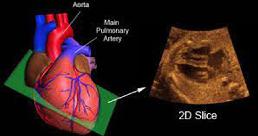
Fetal 2D echo, short for fetal echocardiography, is a specialized ultrasound examination performed during pregnancy to evaluate the structure and function of the fetal heart. This procedure is typically recommended when there is a suspicion of a congenital heart defect or when there are other risk factors present. Using high-frequency sound waves, the fetal echocardiogram provides detailed images of the fetal heart, allowing the healthcare provider to assess the chambers, valves, and blood flow patterns. It helps in identifying abnormalities such as ventricular septal defects, atrial septal defects, tetralogy of Fallot, and other complex cardiac conditions. Fetal 2D echo is a crucial diagnostic tool that aids in early detection, counseling, and planning for the management of potential cardiac issues before or after birth, ensuring optimal care for the baby and providing valuable information for expectant parents and healthcare providers.